CHEst PHysical Examination integrated with UltraSound – Phase (CHEPHEUS1). A survey of Accademia di Ecografia Toracica (AdET)
Keywords:
Chest ultrasound, Early diagnosis, Diagnostic approachAbstract
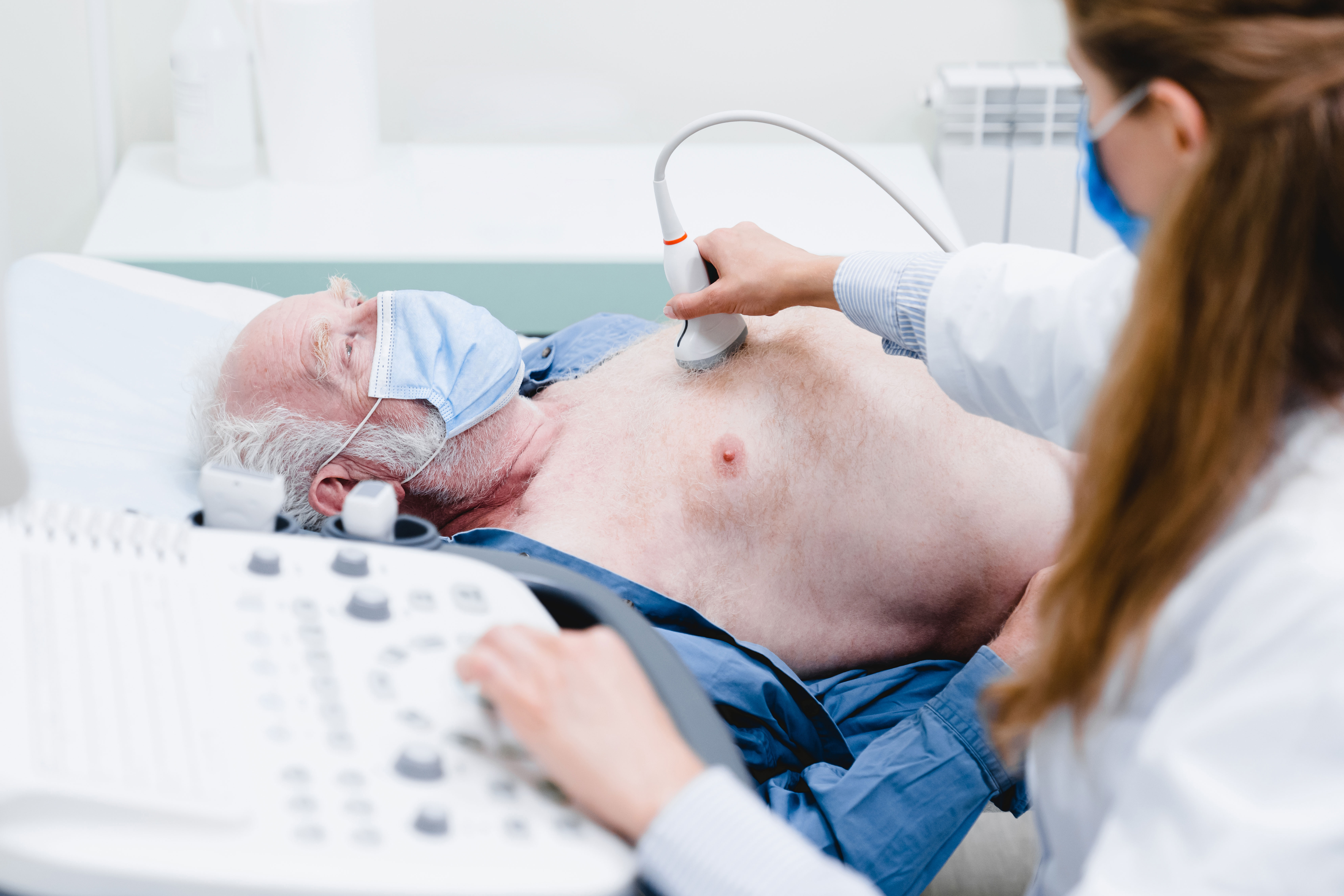
Background: Chest physical exam (CPE) is based on the four pillars of classical semiotics. However, CPE’s sensitivity and specificity are low, and is affected by operators’ skills. The aim of this work was to explore the contribution of chest ultrasound (US) to the traditional CPE.
Methods: For this purpose, a survey was submitted to US users. They were asked to rate the usefulness of classical semiotics and chest US in evaluating each item of CPE pillars. The study was conducted and described according to the STROBE checklist. The study used the freely available online survey cloud-web application (Google Forms, Google Ireland Ltd, Mountain View, CA, USA).
Results: The results showed a tendency to prefer chest US to palpation and percussion, suggesting a possible future approach based on inspection, auscultation and palpatory ultrasound evaluation.
Conclusion: The results of our survey introduce, for the first time, the role of ultrasound as a pillar of physical examination. Our project CHEPHEUS has the aim to study and propose a new way of performing the physical exam in the future.
References
1. Fulton JF. History of medical education. Br Med J 1953;2:457-61.
2. Osler W. The quotable Osler. Silverman ME, Murray TJ, Bryan CS, eds. Philadelphia, PA: American College of Physicians, American Society for Internal Medicine; 2003.
3. Soldati G, Smargiassi A, Mariani A, Inchingolo R. Novel aspects in diagnostic approach to respiratory patients: is it the time for a new semiotics? Multidiscip Respir Med 2017;12:15.
4. Narula J, Chandrashekhar Y, Braunwald E. Time to add a fifth pillar to bedside physical examination: inspection, palpation, percussion, auscultation, and insonation. JAMA Cardiol 2018;3:346-50.
5. Moore CL, Copel JA. Point-of-care ultrasonography. N Engl J Med 2011;364:749-57.
6. Kruisselbrink R, Chan V, Cibinel GA, Abrahamson S, Goffi A. I-AIM (indication, acquisition, interpretation, medical decision-making) framework for point of care lung ultrasound. Anesthesiology 2017;127:568-82.
7. Agricola E, Bove T, Oppizzi M, Marino G, Zangrillo A, Margonato A, et al. Ultrasound comet-tail images: a marker of pulmonary edema: a comparative study with wedge pressure and extravascular lung water. Chest 2005;127:1690-5.
8. Volpicelli G, Skurzak S, Boero E, Carpinteri G, Tengattini M, Stefanone V, et al. Lung ultrasound predicts well extravascular lung water but is of limited usefulness in the prediction of wedge pressure. Anesthesiology 2014;121:320-7.
9. Lichtenstein D, Mézière G, Biderman P, Gepner A, Barrè O. The comet-tail artifact: an ultrasound sign of -alveolar-interstitial syndrome. Am J Respir Crit Care Med 1997;156:1640-6.
10. Jambrik Z, Monti S, Coppola V, Agricola E, Mottola G, Miniati M, et al. Usefulness of ultrasound lung comets as a nonradiologic sign of extravascular lung water. Am J -Cardiol 2004;93:1265-70.
11. Volpicelli G, Mussa A, Garofalo G, Cardinale L, Casoli G, Perotto F, et al. Bedside lung ultrasound in the assessment of alveolar-interstitial syndrome. Am J Emerg Med 2006;24:689-96.
12. Kirkpatrick AW, Sirois M, Laupland KB, Liu D, Rowan K, Ball CG, et al. Hand-held thoracic sonography for detecting post-traumatic pneumothoraces: the extended focused assessment with sonography for trauma (EFAST). J Trauma 2004;57:288-95.
Published
Issue
Section
License
Copyright (c) 2025 The Author(s)

This work is licensed under a Creative Commons Attribution-NonCommercial 4.0 International License.
Mattioli 1885 has chosen to apply the Creative Commons Attribution NonCommercial 4.0 International License (CC BY-NC 4.0) to all manuscripts to be published.
How to Cite